We don’t know if a sperm actually experiences joy when it finally finds the egg, but it does wiggle excitedly.
Patricia A. Martin-DeLeon, a reproductive biologist at the University of Delaware, has witnessed this behaviour many times in her studies of fertility in mice, the closest genetic model to humans (and with a much faster reproductive cycle).
It’s what happens next in the fertilisation process that DeLeon and her team have revealed for the first time, which could help couples struggling with infertility.
“There is communication between the sperm and the fallopian tube that helps prepare the sperm for its big push into the egg,” says DeLeon, who is the Trustees Distinguished Professor of Biological Sciences at UD.
The research, supported by the National Institutes of Health-National Institute of Child Health and Human Development and the Delaware INBRE programme, is published in the Journal of Biological Chemistry. It is one of the top most viewed articles published online this summer under the Membrane Biology affinity group, according to the editorial offices of the American Society for Biochemistry and Molecular Biology.
Understanding what happens in the fertilisation process takes a little walk down biological memory lane, a reminder of nature’s course that led to most of us.
Once the egg is released from an ovary and enters the fallopian tube, the hair-like cilia that line this tiny tube sweep the egg toward the uterus. While in the tube, the egg will either meet the sperm and be fertilised, which must happen within a 12- to 24-hour window of time, or fertilisation will not occur and the egg will dissolve.
DeLeon and her UD team have identified particles in the secretions from the fallopian tube that help the sperm get ready for its all-important drive into the end zone. They termed these bodies “oviductosomes,” from “oviduct,” which is another name for the fallopian tube, and “exosomes,” the teeny sacs found in all the body’s fluids that help cells survive.

Hormones trigger the release of these oviductosomes, which are only 100 nanometers in diameter, or about 10 millionths of an inch wide. The tiny cargo-filled sacs then attach to the sperm like decorations on a Christmas tree before the sperm fuses with the egg.
Once these sacs are in place, they transfer proteins, including a “calcium clearance pump” (scientifically known as Plasma Membrane Ca2+-ATPase 4), to the sperm for use when the time is right.
“This calcium pump is required by the sperm just prior to fertilisation, as well as in the early embryo,” DeLeon says. “The sperm pumps out calcium and takes in hydrogen ions, which seems to give it that last push into the egg, and also is critical to starting the zygote’s life.”
DeLeon and her team made their discovery using the high-powered, three-dimensional super-resolution microscope that UD acquired in 2012. The technology, whose inventors won the Nobel Prize in 2014, can illuminate what’s happening in a cell, right down to a single molecule.
The oviductosomes from a female mouse were pre-labeled with a fluorescent dye and incubated together with the sperm. Within an hour, the oviductosomes were fused to the sperm’s surface. After two to three hours, the oviductosomes continued to accumulate, primarily on the sperm’s head and the midpiece of its tail.
Source: sciencedaily.com
ABUJA: Training Schedule for Basic Life Support BLS, Pediatric Advanced Life Support (PALS), Advanced Cardiovascular Life Support ACLS, First Aid, CPR, AED
PORTHARCOURT: Training Schedule for Basic Life Support BLS, Pediatric Advanced Life Support (PALS), Advanced Cardiovascular Life Support ACLS, First Aid, CPR, AED
LAGOS: Training Schedule for Basic Life Support BLS, Pediatric Advanced Life Support (PALS), Advanced Cardiovascular Life Support ACLS, First Aid, CPR, AED
STOP paying for airtime and electricity, Let your phone pay its bills with ScreenT
You may also like

Comrade Lemii Christian Emerges Chairman of NANNM Rivers State Branch in Historic Victory
PORT HARCOURT, NIGERIA Following the long awaited National Association of N…

FG Supports Robotic Surgery Expansion to Reduce Medical Tourism
Nigeria is taking another step toward strengthening specialised healthcare …
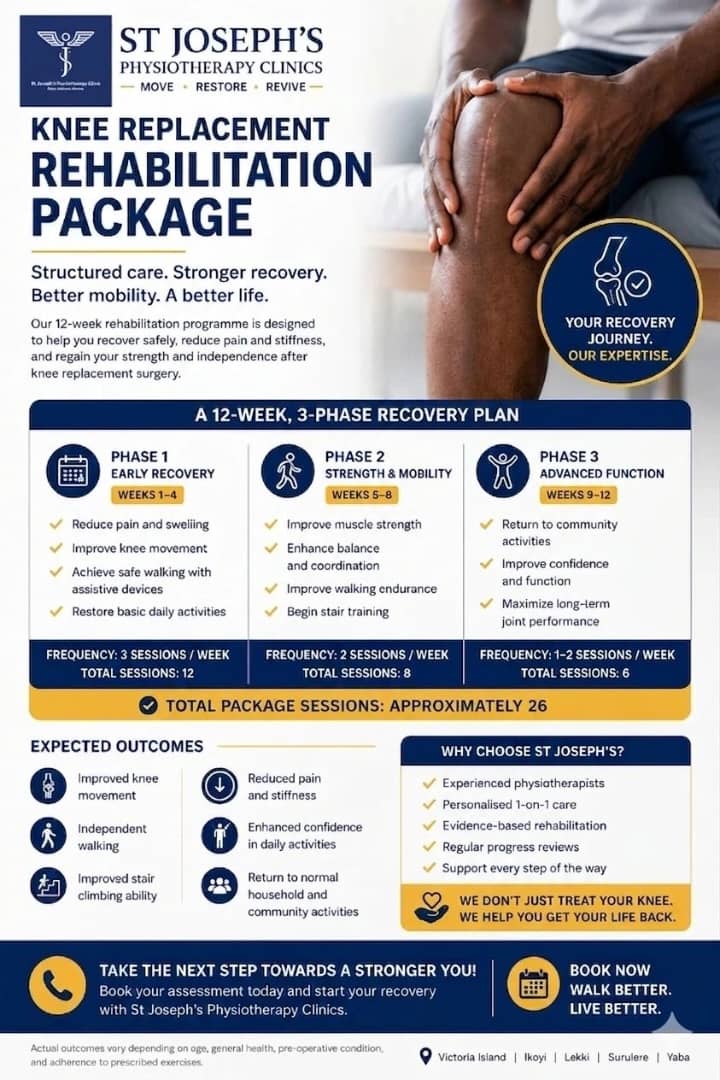
Complete Knee Replacement Rehabilitation Package in Lagos
Knee replacement surgery is a major step toward pain relief and improved mo…
NAMDA threatens strike over unpaid medical lecturers’ allowances
The Nigerian Association of Medical and Dental Academics (NAMDA) has warned…
Resident Doctors Signal End to Strike Threat After High-Level Talks with Gbajabiamila
The threat of a nationwide indefinite strike by the Nigerian Association of…