. Anatomage Table shows images of life-sized bodies and insides at any angle
. Students can 'virtually dissect' the cadaver at the touch of a screen
. Organs, veins, nerves and tissue can be removed and put back in again
. Device to be introduced for teaching medical students 'where appropriate' at Edinburgh University
A hi-tech 'virtual cadaver' is being made available for teaching - so students can investigate the human anatomy at the touch of a screen.
The device, at The University of Edinburgh’s School of Anatomy, will enable medical students to examine the human body by ‘virtually dissecting it’. Dubbed the Anatomage Table, it shows life-sized male and female bodies created from CT scans, allowing the cadaver to be seen from front to back, side to side and upside down.

Virtual cadaver: The Anatomage Table will enable students to 'virtually dissect' a human body

Life-sized: Images of male and female bodies have been created from CT scans, allowing the cadaver to be seen from front to back, side-to-side and upside down
It means the body and the relationship of structures beneath the skin can be seen in great detail – and electronically.
Unlike the dissection of a real cadaver, in which body parts can only be removed, students can add or remove organs, veins, arteries, nerves or tissue by touching the table - enabling them to see how one part relates to another.
Gordon Findlater, Professor of Translational Anatomy, said: ‘The beauty of the Anatomage Table is that you can rotate and view the body in all three planes in a unique 3D experience.
‘Although it will never, I believe, replace the experience of dissecting and handling a real cadaver, it will allow students to handle a virtual cadaver without all the legislation that accompanies the use of real cadavers.

Researchers say it is possible to import personalised scans to the device, which can then be recreated into virtual cadavers and used in specially designed teaching packages

Students can add or remove organs, veins, arteries, nerves or tissue by touching the table - enabling them to see how one part relates to another

Handy: Body parts can be seen in great detail thanks to the touch-screen teaching tool
‘So far we have received a lot of good feedback from the students and surgeons who have tested it out.’
Researchers say it is possible to import personalised scans to the device, which can then be recreated into virtual cadavers and used in specially designed teaching packages.
The device will be introduced into formal teaching sessions ‘where appropriate’ and will also be put on display for members of the public.
By Julian Robinson
georgianewsday
ABUJA: Training Schedule for Basic Life Support BLS, Pediatric Advanced Life Support (PALS), Advanced Cardiovascular Life Support ACLS, First Aid, CPR, AED
PORTHARCOURT: Training Schedule for Basic Life Support BLS, Pediatric Advanced Life Support (PALS), Advanced Cardiovascular Life Support ACLS, First Aid, CPR, AED
LAGOS: Training Schedule for Basic Life Support BLS, Pediatric Advanced Life Support (PALS), Advanced Cardiovascular Life Support ACLS, First Aid, CPR, AED
STOP paying for airtime and electricity, Let your phone pay its bills with ScreenT
You may also like
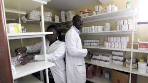
PSN Raises Alarm as 3 Million Illegal Drug Outlets Threaten Public Safety
The Pharmaceutical Society of Nigeria, PSN, in Lagos State yesterday warned…

Agencies move to strengthen early warning systems in Nigeria’s healthcare sector
Key government and international agencies have intensified efforts to stren…
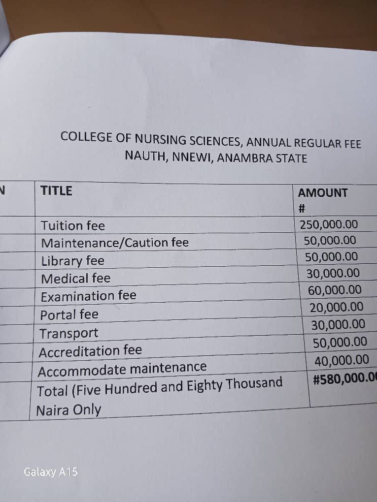
NAUTH management moves to resolve tuition hike protest, promises dialogue with students
The management of Nnamdi Azikiwe University Teaching Hospital (NAUTH) has p…

Child health outcomes improve as facility-based maternal deaths decline in 2025 FG report
The Federal Government has reported significant improvements in maternal, n…
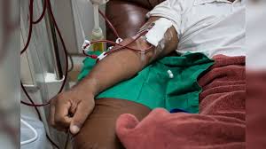
World Kidney Day: Caregivers groan as dialysis hits N80,000 per session, transplant N30m
As the world marks World Kidney Day, caregivers and health experts in Niger…